Shedding New Light
on Imaging Core
MHIR NEWS
March 27, 2018
Shedding New Light On Imaging Core
MHIR’s Small Animal Imaging Facility Unique to Area, Key to Research
The Small Animal Imaging Facility provides magnetic resonance imaging (MRI) and high-resolution ultrasound imaging. As with a clinical MRI for humans, this equipment uses a powerful magnetic field, radio frequency pulses and a computer to produce detailed pictures of soft tissues, including virtually all organs and other internal body structures of a small animal. At MaineHealth Institute for Research (MHIR), this non-invasive equipment is utilized to gather both structural and functional information in mice to further research and it is one of thirteen Research Core Facilities and Services at MHIR.

Dr. Ilka Pinz preparing a sample to be scanned in the BRUKER PharmaScan MR imager.
The Small Animal Imaging Facility equipment includes a BRUKER PharmaScan 7T, 300 MHz imager with a resolution of 90 µm, which is an extremely powerful super-cooled magnet. While it is commonly used at MHIR to facilitate the research of MHIR investigators, all of this
equipment is available for external investigators as well. The animal facility provides non-barrier housing for study animals from outside institutions. According to Ilka Pinz, PhD, Faculty Scientist at MHIR and head of the MRI Imaging Core, “It’s exciting what we are able to provide in addition to the typical services of an MRI – getting a closer look at nanoparticles or nanostructures and their in vivo effects as well as any kind of small animal disease model critical to biomedical research.”
While there are other facilities like this in New England, the Imaging Core at MHIR is unique to the immediate region– “We are the only small animal imaging facility in Maine, and even though the equipment is most commonly used for mice, it can also be used for rats, crabs, and even fresh water fish,” said Dr. Pinz.
A list of MRI services available through MHIR include:
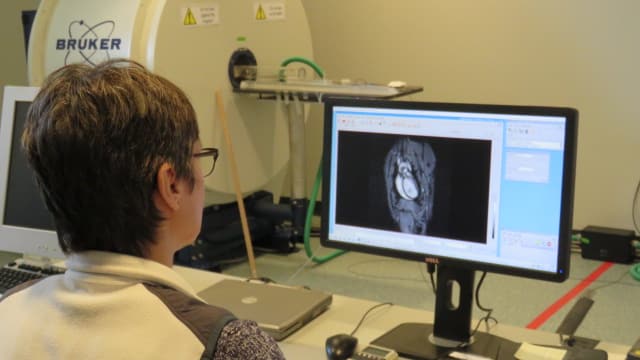
Dr. Pinz reviewing and measuring cardiac MR images at the MR workstation.
• Morphological images of all organs.
• Magnetic resonance angiography, non-contrast and contrast enhanced.
• Proton spectroscopy, global and localized (for example: assessment of water: fat ratios in bone marrow).
• Cardiac imaging, diastolic and systolic dimensions of the ventricles, cardiac movies, late gadolinium, enhancement of cardiac infarcts, cardiac scar evaluation and size determination.
• In-utero assessment of embryo development: determination of embryonic defects and determination of the time point for the loss/resorption of transgenic embryos. Capable of high-resolution embryo imaging with ~27 µm spatial resolution.
• Targeted contrast enhanced imaging using your specific antibody that is conjugated to gadolinium.
The Image Core also has high-resolution ultrasound equipment. Currently, the ultrasound (VEVO2100, VisualSonics) imaging is within MHIR’s barrier facility and is only available for mice from approved animal vendors. Services provided include: anatomical/morphological imaging, tumor volume determination, echocardiography, targeted and non-targeted microbubble imaging, and blood flow quantification.
Moving forward Dr. Pinz hopes to see more outside investigators utilize the facility— “The challenge is getting the word out to researchers about this Imaging Core’s capabilities and the state-of-the art services we can provide to help advance bio-medical research,” said Dr. Pinz.
For more information about the Imaging Core at MHIR, services, and pricing please contact Dr. Pinz at pinzi@mainehealth.org.